Acoustic 3D Printing
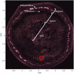
Acoustic 3D printing uses focused ultrasound to create 3D objects. The focused ultrasonic energy causes localized heating which is used to solidify materials. The unique feature of this fabrication method is that ultrasound can be transmitted through objects and obstacles, same as it is done for medical imaging and treatments. This allows the printing inside of existing structures and the combination of various materials. This is an advantage over optical 3D printing methods where the structure must be transparent for light.
In 2025, Dr. Martin Weber has received research funding from the Jane and Aatos Erkko Foundation [https://jaes.fi/en/frontpage/] which enables him to expand this research.
Highlights:
Weber, M., Nikolaev, D., Koskenniemi, M. et al. Fabrication of poly (ε-caprolactone) 3D scaffolds with controllable porosity using ultrasound. Sci Rep 15, 23415 (2025). https://doi.org/10.1038/s41598-025-06818-9
Contact:
Dr. Martin Weber
martin.weber[at]helsinki.fi
Levitator
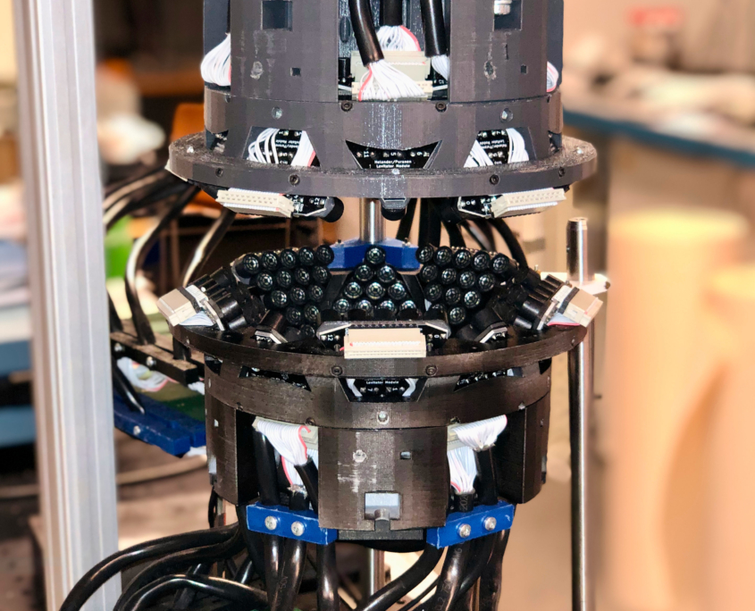
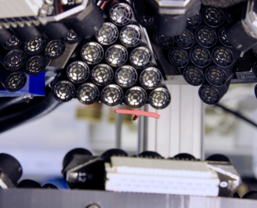
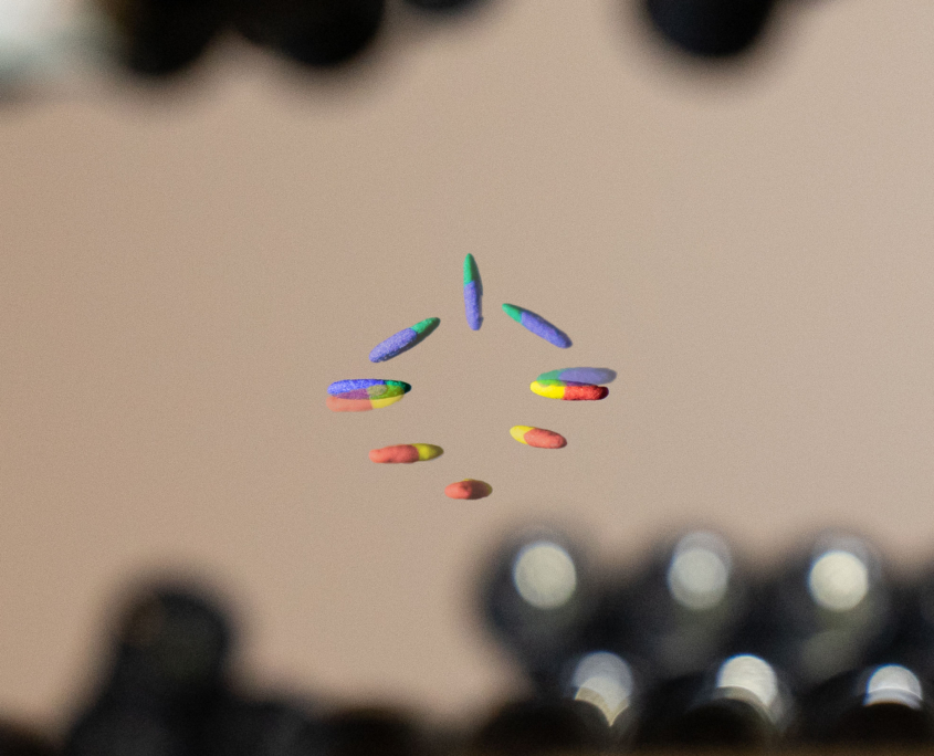
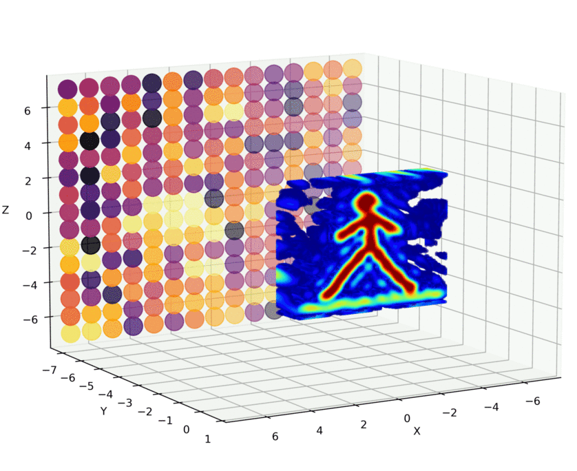
Acoustic sound waves create acoustic sound pressure that can exert forces on the objects and suspend them against gravity in mid-air, enabling acoustic levitation. We built the levitator that consist of 450 ultrasonic transducers (f = 40 kHz, diameter = 1 cm) arranged into 2 hemispheres. Each transducers’ amplitude and phase can be controlled individually that allow us to design complex pressure profiles to levitate cm-sized objects or to create 3D holograms.
Within the project we are developing:
- Levitator electronics with control software, design of modular units
- Optimisation algorithms for solving control parameters (levitators’ amplitudes and phases)
- Dynamics of the levitating objects with FEM simulations
- Artificial Intelligence tools to control the levitation process
Highlights:
- “Omnidirectional microscopy by ultrasonic sample control“ Appl. Phys. Lett. 116, 194101 (2020)
P. Helander, T. Puranen, A. Meriläinen, G. Maconi, A. Penttilä, M. Gritsevich, I. Kassamakov, A. Salmi, K. Muinonen, and E. Hæggström. - “Multifrequency acoustic levitation“ in IEEE International Ultrasonics Symposium, 916 (2019)
T. Puranen, P. Helander, A. Meriläinen, G. Maconi, A. Penttilä, M. Gritsevich, I. Kassamakov, A. Salmi, K. Muinonen, and E. Haeggström.
Contact:
Dr. Denys Iablonskyi
denys.iablonskyi[at]helsinki.fi
Ultrasound-Enhanced Electrospinning
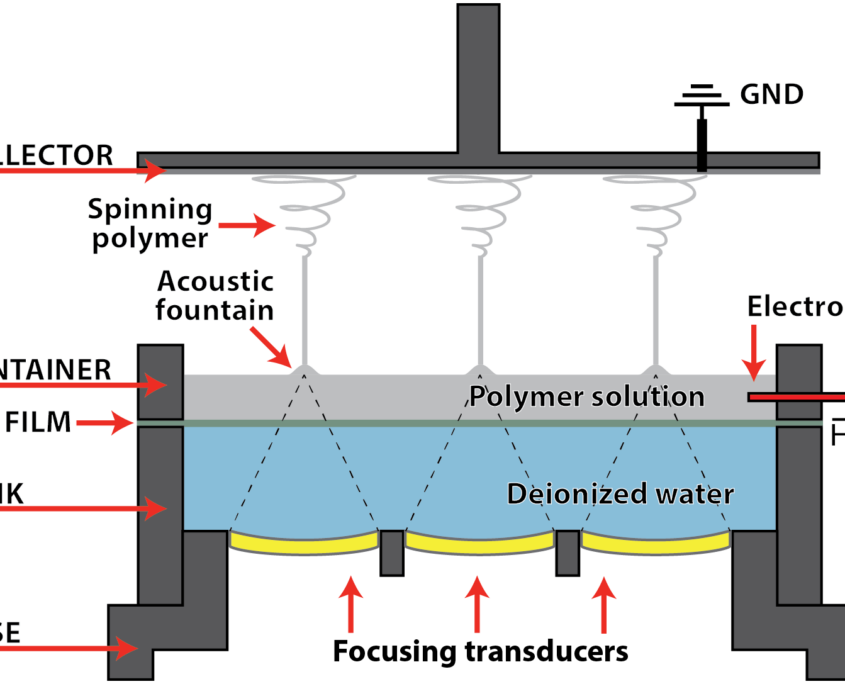
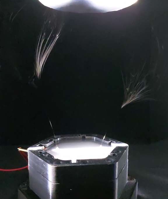
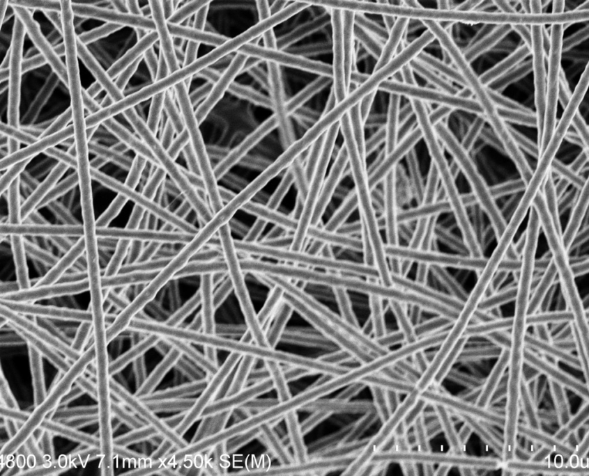
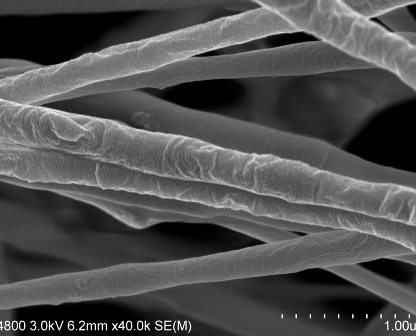
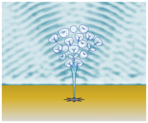
Ultrasound-enhanced electrospinning (USES) is an alternative to traditional electrospinning processes for the producing nanoscale polymer fibers. The system uses focused ultrasound creating a surface protrusion (acoustic fountain) to initiate electrospinning. The method eliminates disadvantages of the traditional electrospinning process and adds the ability to control fiber diameter and morphology, which can be modified by varying ultrasound parameters (frequency, amplitude, pulse repetition frequency, cycles per pulse).
The following works are carried out within the framework of the project:
- Simulation of the effect of ultrasound parameters on the process of fiber formation using Finite Element Method (FEM)
- Conducting experimental work to reveal the influence of experimental setup parameters, ultrasound parameters, polymer composition on the process of fiber formation, as well as obtaining fibers mats for subsequent analysis
- Analysis of the formed fibers using electron microscopy, X-ray diffraction and other methods
- Scaling the system and optimizing its parameters to realize the possibility of industrial application of the method
1) “Ultrasound-enhanced electrospinning“, Scientific Reports (2018), H. J. Nieminen et al.
2) “Comparison of Traditional and Ultrasound-Enhanced Electrospinning in Fabricating Nanofibrous Drug Delivery Systems“, Pharmaceutics (2019), E. Hakkarainen et al.
Contact:
Dr. Dmitry Nikolaev
dmitry.nikolaev[at]helsinki.fi
Non-Destructive Testing
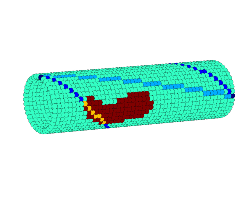
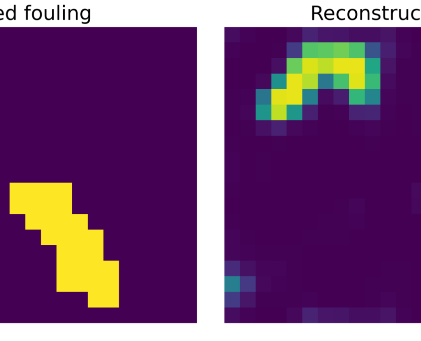
Ultrasonic guided waves (UGWs) can propagate over long distances with little attenuation and are suitable for non-destructive evaluation of a large area. However, localizing fouling inside the closed systems (e.g., pipes, engines) remains to be challenging. Here we propose to use UGWs that propagate along the pipe circumference along multiple helical paths, thus, providing denser structural information without increasing the number of sensors. When the path crosses fouled area the UGWs propagation properties change, generating unique features into each helical wave. Such aggregate information together with the known structural geometry are then used to fit a latent variable Gaussian process to recover the fouling distribution map in a probabilistic manner.
Contact:
Dr. Denys Iablonskyi
denys.iablonskyi[at]helsinki.fi
Localized Material Removal with Ultrasound
Ultrasound Imaging Mass Spectrometry (UIMAS)
UIMAS is a sampling method that combines ultrasound and mass spectrometry. Ultrasound is first used to image a sample surface. If something interesting is found, material is locally removed with HIFU-induced cavitation (sampling size 40–400 µm) and the particles are transported to a mass spectrometer for chemical analysis. The video shows a marker ink sample being removed from a glass plate. We are interested in applying this method for biological samples, where mechanical imaging can be combined with chemical analysis.
Highlights:
- Sillanpää, J. Hyvönen, J. Mäkinen, A. Holmström, T. Pudas, P. Lassila, R. Lepistö, A. Kuronen, T. Kotiaho, E. Hæggström, and A. Salmi, “Ultrasound-based surface sampling in immersion for mass spectrometry,” Journal of Applied Physics, 134(10), 2023, Art no. 104901, doi: https://doi.org/10.1063/5.0157705.
Contact:
M.Sc. Axi Holmström
e-mail: axi.holmstrom[at]helsinki.fi
CESAM – Coded Excitation Scanning Acoustic Microscopy
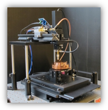
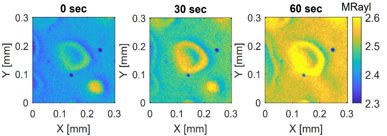
Our scanning acoustic microscope employs coded excitation to achieve large bandwidth and high signal-to-noise ratio (SNR). These are required for imaging biological samples with low contrast (soft tissue has an acoustic impedance that is close to the impedance of water) and with samples that have layered structures (multiple echoes arriving closely in time). We are developing the device to achieve higher SNR and lower measurement time to study dynamical systems. One of our current research projects is related to bone re-ossification post fracture.
Contact:
Dr. Martin Weber
martin.weber[at]helsinki.fi
Laser ultrasonics
High-energy laser pulses can be used to generate ultrasonic waves. They deposit energy into a material which causes thermal expansion on a nanosecond time scale. That leads to a sudden change of the mechanical stress in the material and generates an ultrasonic wave.
The detection of an ultrasonic wave can be done optically, too. For example, a laser interferometer measures the displacement of the material’s surface.
We are developing laser ultrasonic and ultrasonic imaging methods in collaboration with several industry partners. Applications range from ultrasonic quality control of materials to laser ultrasonic tomography.
Highlights:
1. “Defect localization by an extended laser source on a hemisphere” Scientific Reports volume 11, 15191 (2021), https://doi.org/10.1038/s41598-021-94084-w
Daniel Veira Canle, Joni Mäkinen, Richard Blomqvist, Maria Gritsevich, Ari Salmi & Edward Hæggström
2. “Contactless Damage Evaluation of a Rotating Carbon Fiber Propeller” 2019 IEEE International Ultrasonics Symposium (IUS) https://doi.org/10.1109/ULTSYM.2019.8925618
Daniel Veira Canle, Joni Mäkinen, Petri Lassila, Maria Gritsevich, Ari Salmi, Edward Hæggström
Contact:
Dr. Martin Weber
martin.weber[at]helsinki.fi
Focused UltraSound for Urban MINing (FUSUMIN)
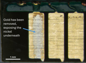
We use focused ultrasound to 1) image a sample (low power) and 2) create cavitation bubbles, which locally erode material from a desired spot on the sample surface (high power). Eroded particles are freed into the water bath and can then be collected. The method has been applied to two projects, Ultrasound Imaging Mass Spectrometry (UIMAS) and Focused Ultrasound for Urban Mining (FUSUMIN):
Contact:
M.Sc. Axi Holmström
e-mail: axi.holmstrom[at]helsinki.fi
Contact details
Electronics Research Laboratory
Department of Physics
P. O. Box 64
(Gustaf Hällströmin katu 2)
FIN-00014
UNIVERSITY OF HELSINKI
FINLAND
Google map
Tel. +358-2-941 50693
